How to Mix Lyophilized Powder: Reconstitute in Simple Steps
Disclaimer: The information provided in this article is for educational and informational purposes only. It is not intended as medical advice, diagnosis, or treatment. Products and compounds referenced are for research use only and are not approved for human or veterinary consumption. Always consult a qualified professional regarding health or medical decisions.
Understanding Lyophilization and Reconstitution Principles
Understanding how to mix lyophilized powder begins with letting the vial reach room temperature, calculating the required diluent volume for your target concentration, slowly introducing the diluent along the inner glass wall, and gently swirling until the solution is clear. Lyophilization, commonly known as cryodesiccation or freeze-drying, is a low-temperature dehydration method designed to preserve heat-sensitive compounds. By removing moisture through sublimation—where ice converts directly into vapor under vacuum without passing through an intermediate liquid phase—the process protects delicate chemical structures. A comprehensive scientific background on freeze drying demonstrates how this thermodynamic pathway prevents the thermal denaturation typically caused by conventional heat drying.
The freeze-drying cycle begins with freezing the compound below its triple point, followed by primary drying (sublimation), which strips away approximately 95% of the formulation’s water. A subsequent secondary drying phase desorbs chemically bound water, leaving a finished cake with an exceptionally low residual moisture level of just 1% to 4%.
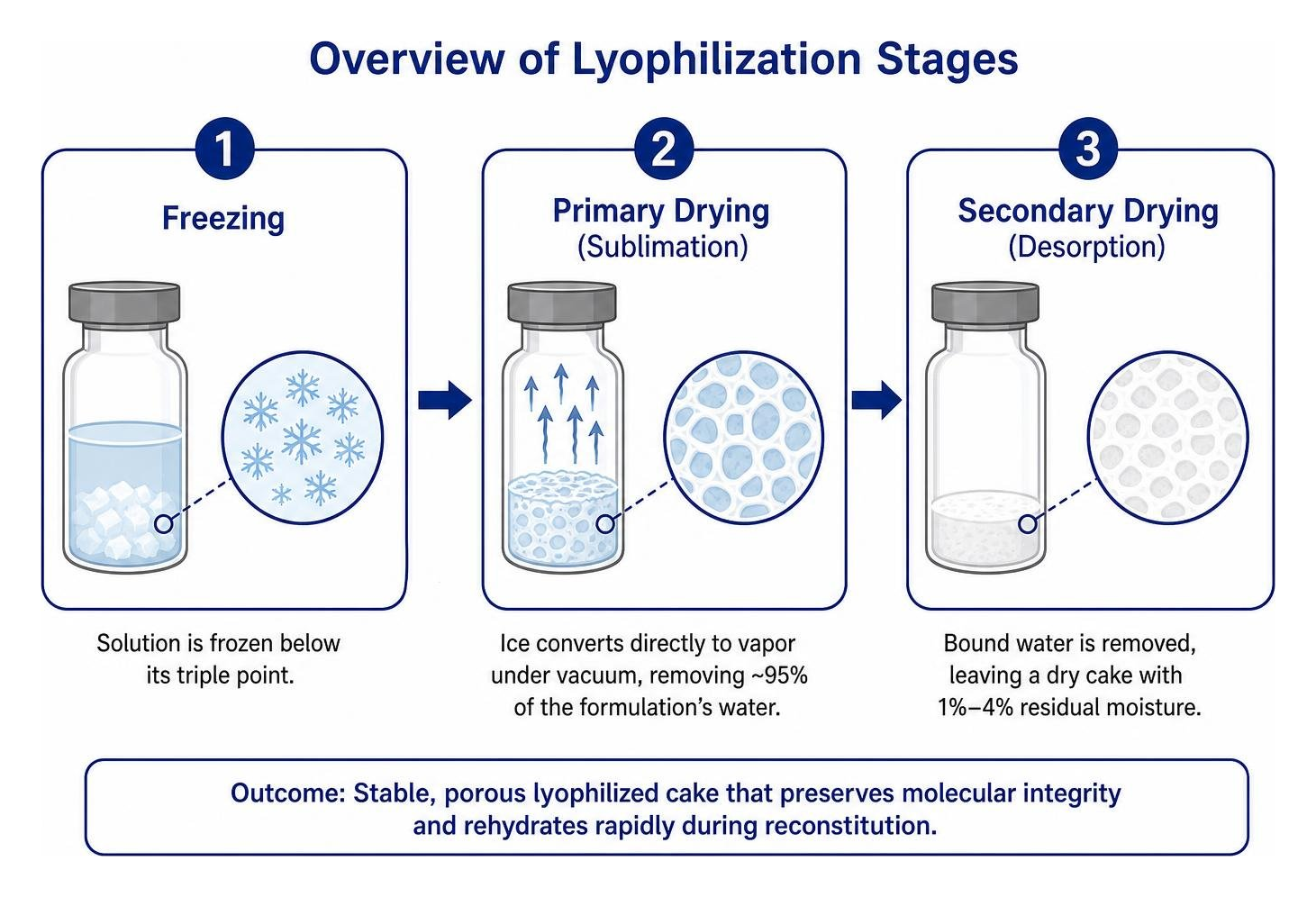
This intricate dehydration mechanism forms a microscopic, sponge-like lattice across the solid cake. Understanding why research peptides are typically lyophilized clarifies why this specific structure is essential: those microscopic pores allow reconstituting solvents to penetrate rapidly, ensuring smooth rehydration while safeguarding molecular integrity. These porous cakes maintain structural stability across prolonged experimental storage.
Types of Lyophilized Formulations and Diluent Requirements
Different scientific compounds require specific solvent environments depending on their molecular charge, hydrophobicity, and intended experimental workflow. High-molecular-weight proteins, synthetic peptides, and complex biological matrices each exhibit distinct chemical behaviors when reintroduced to liquid media.
| Formulation Type | Primary Diluents | Stability Window (Reconstituted) | Recommended Research Application |
|---|---|---|---|
| Synthetic Research Peptides | Bacteriostatic Water (0.9% Benzyl Alcohol), Sterile Water | 21–28 Days at 2°C–8°C (with preservative) | Multi-aliquot laboratory protocols, analytical assays |
| Monoclonal Antibodies & Enzymes | Sterile Water, Specific Formulation Buffers (Tris-HCl, PBS) | 24–48 Hours at 2°C–8°C (Preservative-Free) | Enzymatic assays, cell-free binding analysis |
| Biological Matrix / Plasma | Sterile Distilled Water, 0.9% Sodium Chloride | Single analytical session | In vitro coagulation research, emergency trauma models |
The efficiency of lyophilized matrices is widely demonstrated across biological literature. For example, published clinical trial evidence on lyophilized plasma reconstitution (Garrigue et al., 2018) highlights how freeze-dried biological formulations dissolve rapidly—often within minutes—without requiring lengthy thermal thawing cycles, preserving critical factors for experimental analysis.
Selecting the Appropriate Diluent
Selecting the proper liquid base is crucial when preparing your research solutions. Using an incompatible solvent can cause immediate precipitation, peptide cleavage, or rapid bacterial contamination.
- Bacteriostatic Water: Sterile water containing 0.9% benzyl alcohol as an antimicrobial preservative. It is the preferred diluent for multi-use research containers because it prevents microbial proliferation over several weeks of refrigerated storage.
- Sterile Water: High-purity, endotoxin-free water without added antimicrobial agents. Ideal for single-use experimental runs, sensitive cellular cultures, or assays where benzyl alcohol might interfere with enzymatic activity. Understanding the sterile water versus bacteriostatic water differences ensures researchers select the correct vehicle for their protocol.
- Normal Saline (0.9% NaCl): An isotonic solution used when maintaining specific osmotic pressure is vital for the stability of sensitive proteins.
- Specialized Buffers (Tris-HCl, Dilute Acids): Certain proteins and enzymes require strictly controlled pH ranges. For instance, specific enzymes demand Tris buffers with calcium cofactors, while acidic solutions (such as 0.01 N HCl) are necessary to prevent rapid aggregation during the reconstitution of collagen or specialized basic peptides.
Maintaining proper bacteriostatic water storage protocols is equally essential to prevent solvent degradation before introducing it to the lyophilized cake.
Equipment Setup and Sterility Protocols
Aseptic technique is vital when reconstituting laboratory reagents. Because reconstituted solutions contain no immune defenses, environmental microorganisms introduced during preparation can rapidly degrade the target sequence and invalidate experimental outcomes.
Begin by creating a sterile research environment inside a laminar flow hood or a clean, sanitized bench space free of air turbulence. Thoroughly clean all working surfaces with 70% isopropyl alcohol and let the area air-dry completely. For a full breakdown of workspace setup and sterile tools, refer to our comprehensive peptide supplies guide 2026.
Essential Supplies for Mixing Reagents
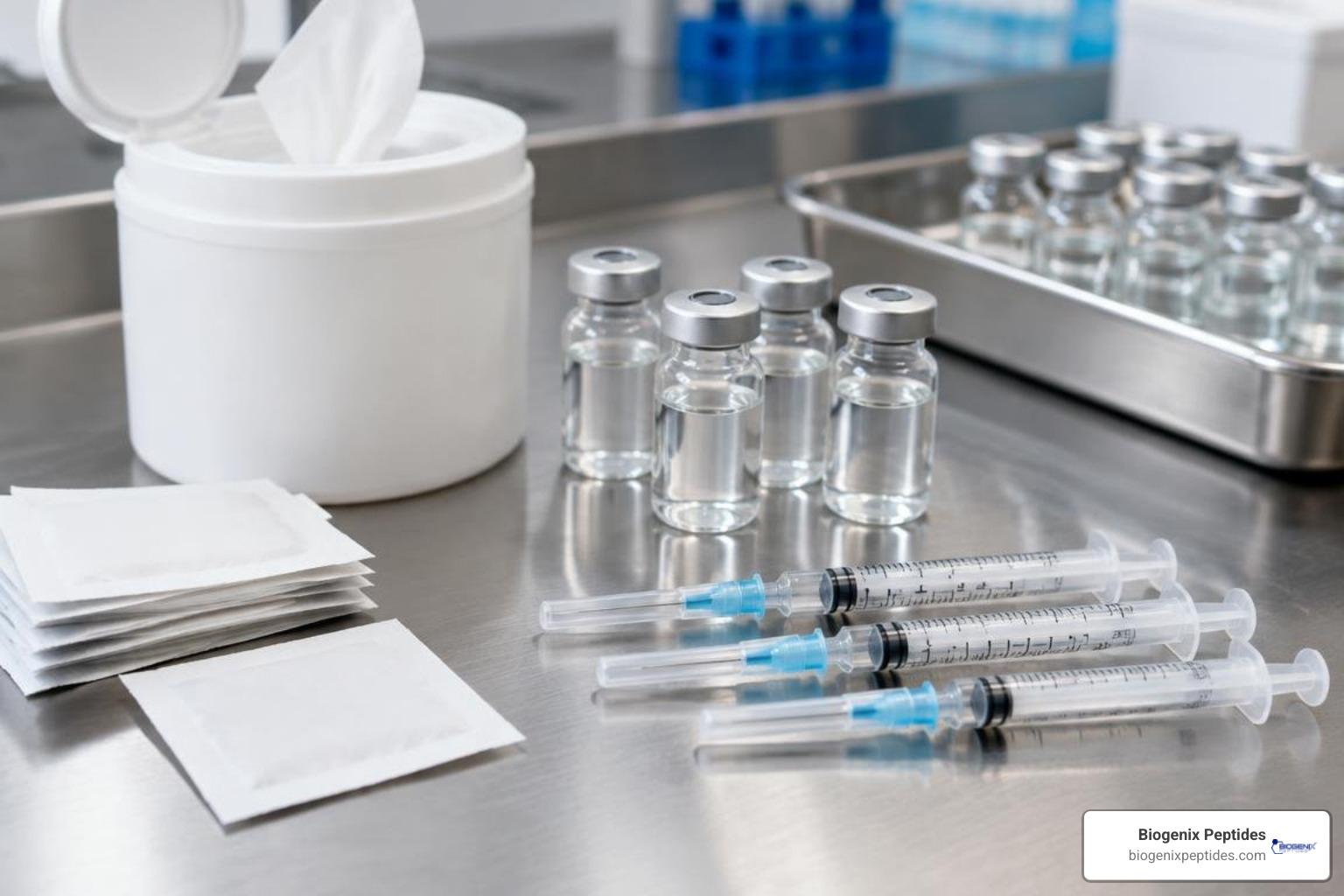
Before unsealing containers, gather all necessary labware to maintain continuous workflow sterility:
- Lyophilized compound vial (equilibrated to ambient room temperature)
- Appropriate reconstitution diluent (e.g., bacteriostatic water)
- Single-use, disposable syringes with ultra-fine calibration markings
- Mixing needles (typically 20G to 22G for solvent transfer, or integrated fine needles)
- Individual 70% isopropyl alcohol prep pads
- Dedicated sharps biohazard container
Using certified sterile syringes for research reconstitution guarantees accurate volume delivery while minimizing particulate contamination from low-grade manufacturing materials.
Calculating Diluent Volumes and Target Concentrations
Accurate laboratory concentration math is essential for reproducible scientific trials. Adding arbitrary liquid volumes compromises experimental precision. The foundational formula for calculating final concentration is:
$$text{Concentration (mg/mL)} = frac{text{Total Mass of Reagent (mg)}}{text{Volume of Diluent Added (mL)}}$$
For example, if you reconstitute a 5 mg vial of lyophilized peptide with 2 mL of bacteriostatic water:
$$text{Concentration} = frac{5text{ mg}}{2text{ mL}} = 2.5text{ mg/mL}$$
To achieve a round concentration that simplifies experimental measurements, consult our bacteriostatic water peptide ratio guidelines or design tailored parameters using our guide to custom peptide dilution ratios.
Step-by-Step Reconstitution Protocol
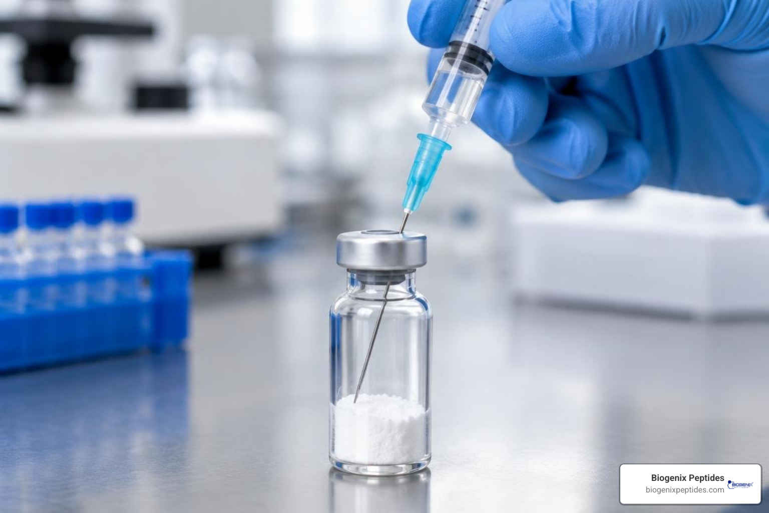
Handling delicate peptide chains and protein complexes requires careful mechanical technique. Rushing the process or creating turbulence can permanently disrupt delicate secondary and tertiary molecular folds. Following a validated sequence for reconstituting lyophilized peptides step by step guarantees maximal yield and biological activity.
Step-by-Step Reconstitution Process
- Temperature Equilibration: Remove the lyophilized vial from cold storage (-20°C or 2°C–8°C) and let it reach room temperature for 15–20 minutes. Opening a cold vial exposes the dry cake to atmospheric humidity, causing instant condensation that can degrade delicate compounds.
- Cap Disinfection: Pop off the plastic flip-off seal from both the reagent vial and the diluent container. Firmly swab both rubber stoppers with an alcohol wipe using a single-direction motion. Let the stoppers air dry for 30 seconds.
- Aspirate Diluent: Draw atmospheric air into the syringe equal to your intended diluent volume. Insert the needle into the diluent vial, invert the bottle, depress the plunger to equalize internal pressure, and smoothly withdraw the measured volume of liquid.
- Controlled Liquid Transfer: Insert the needle through the center of the reagent vial’s rubber septum at a 45-degree angle. Direct the needle tip toward the inner glass wall rather than spraying directly onto the freeze-dried cake.

- Manage the Vacuum: Lyophilized vials frequently maintain an internal negative pressure vacuum. Do not let the diluent slam violently into the cake. Control the syringe plunger with your thumb, allowing the solvent to drip slowly down the glass wall.
- Gentle Dissolution: Once the diluent is transferred, withdraw the syringe. Gently swirl the vial in circular motions or slowly invert it end-over-end. Never shake the vial vigorously.
- Resting Phase: Allow the vial to rest undisturbed at room temperature for 5 to 15 minutes to ensure complete hydration of all microscopic particles.
Practical insights from published practical guidance in lyophilized protein development demonstrate that managing internal headspace vacuum and avoiding aggressive agitation directly minimizes protein aggregation. For additional background on protecting peptide integrity during fluid transfer, review our guide to stopping peptide denaturation.
Handling Vials, Dual-Chamber Syringes, and Specialized Containers
While single glass vials are standard across most research setups, specialized container formats require modified handling:
- Standard Septum Vials: Always pierce the exact center circle of the chlorobutyl stopper to avoid coring rubber particles into the research solution.
- Dual-Chamber Systems: Dual-chamber designs separate the lyophilized cake from the diluent using an internal middle stopper. Depressing the plunger pushes the middle stopper into an internal bypass channel, allowing solvent to flow into the powder chamber. Rotate slowly to mix without creating foam.
- Headspace Pressure Balancing: If high internal negative pressure causes the rubber stopper to indent inward, insert a dry, sterile needle (without a syringe) briefly through the septum to vent the vial and equalize atmospheric pressure.
Quality Inspection and Troubleshooting
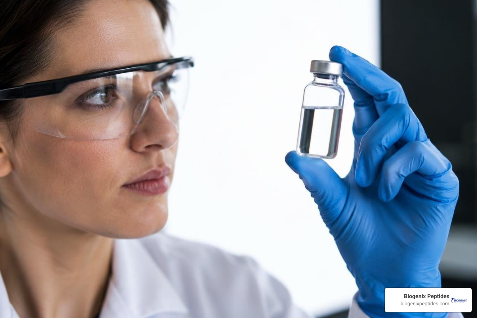
Visual inspection is an essential quality control step following reconstitution. Hold the vial against a bright white background, followed by a dark background, to inspect for clarity, color consistency, and particulates.
A successfully reconstituted solution should appear clear, transparent, and completely free of visible flakes, filaments, or persistent clouds. If the solution shows persistent haziness, review our comprehensive ultimate guide to handling lyophilized powder to pinpoint potential mixing errors.
Common Mistakes and How to Avoid Them
- Vigorous Shaking: Shaking generates high shear stress at the air-water interface, resulting in irreversible protein denaturation and dense foam formation. Always swirl gently.
- Direct High-Velocity Jetting: Allowing diluent to spray directly onto the cake creates insoluble clumps. Always run solvent along the inner glass wall.
- Skipping Temperature Equilibration: Reconstituting a cold vial causes moisture condensation, creating aggregates.
- Selecting the Wrong Diluent: Using plain saline instead of sterile water (or vice versa) can alter ionic strength and cause immediate precipitation.
Reviewing the most common mistakes in peptide research helps laboratory teams prevent these costly protocol errors.
Troubleshooting Incomplete Dissolution and Aggregates
If your lyophilized powder does not dissolve completely within 10 minutes, apply these structured troubleshooting steps:
- Extended Hydration Rest: Let the vial stand undisturbed at ambient temperature for an additional 15 to 20 minutes. Many high-concentration peptide matrices dissolve slowly.
- Gentle Rolling: Roll the vial between the palms of your hands to warm the solution slightly and promote solubilization without introducing shear stress.
- Mild Thermal Incubation: For stubborn compounds (such as hydrophobic sequences), place the vial in a 25°C to 30°C water bath for 5 minutes.
- pH Adjustment for Hydrophobic Peptides: Strongly basic or hydrophobic peptides may require a drop of sterile 0.1% acetic acid to aid dissolution before bringing to volume with water. Highly acidic compounds may benefit from a drop of sterile dilute ammonium hydroxide.
Storage Guidelines and Post-Reconstitution Stability
Once reconstituted, the compound’s protective dry state is gone, making the liquid solution vulnerable to chemical degradation via hydrolysis, deamidation, and oxidation.
Store all reconstituted solutions between 2°C and 8°C (36°F to 46°F). Never store active research solutions in the door of a laboratory refrigerator, where frequent opening causes temperature fluctuations; instead, place them on a stable interior shelf. Discover practical cold storage secrets for reconstituted peptides to maintain peptide stability over time.
Refrigeration and Container Handling
Reconstituted solutions prepared with bacteriostatic water containing 0.9% benzyl alcohol generally remain stable for 21 to 28 days under strict refrigeration. Conversely, solutions reconstituted with preservative-free sterile water must be used immediately or discarded within 24 hours.
For precise storage timelines across various compounds, consult our resource on how long reconstituted peptides last. Protect all peptide solutions from direct ultraviolet light by wrapping vials in aluminum foil or storing them in light-blocking containers.
Frequently Asked Questions
What is the best diluent for mixing lyophilized powder?
For multi-aliquot research requiring extended refrigeration, bacteriostatic water containing 0.9% benzyl alcohol is typically the preferred diluent because it inhibits microbial growth. For immediate, single-use assays or sensitive cell culture applications where preservatives might interfere with cell viability, sterile water is standard. Always consult the specific product certificate of analysis for compound-specific solvent recommendations.
Can you shake a vial to mix lyophilized powder faster?
No. Shaking subjects delicate peptide chains and protein structures to mechanical shear stress, breaking hydrogen bonds and causing irreversible structural denaturation. Shaking also introduces air bubbles and persistent foam that traps the compound, leading to inaccurate volume measurements. Always roll or swirl the vial gently.
How do I know if the lyophilized powder is completely dissolved?
A fully reconstituted compound yields an optically clear solution that matches the viscosity of water, free from floating particulates, cloudiness, or suspended gelatinous matter when held up to a bright light source. If haziness persists after 20 minutes of gentle swirling and resting, the compound may require solvent pH adjustment or have formed irreversible aggregates.
Summary of Best Practices
Mastering laboratory reconstitution is essential for protecting the molecular stability of research peptides and biological matrices. By adhering to strict aseptic protocols, calculating exact diluent volumes, directing solvent along the inner glass wall, and swirling gently without agitation, you preserve the structural integrity of your compounds.
For researchers seeking high-purity research materials and detailed handling protocols, explore our foolproof method to reconstitute lyophilized peptides at BioGenix Peptides to support your laboratory workflows.
References
Cheng Y, Duong HTT, Hu Q, Shameem M, Tang XC. “Practical advice in the development of a lyophilized protein drug product..” Antibody therapeutics, 2025. PMID 39839910. Garrigue D, Godier A, Glacet A, Labreuche J, Kipnis E, Paris C, Duhamel A, Resch E, Bauters A, Machuron F, Renom P, Goldstein P, Tavernier B, Sailliol A, Susen S. “French lyophilized plasma versus fresh frozen plasma for the initial management of trauma-induced coagulopathy: a randomized open-label trial..” Journal of thrombosis and haemostasis : JTH, 2018. PMID 29274254.