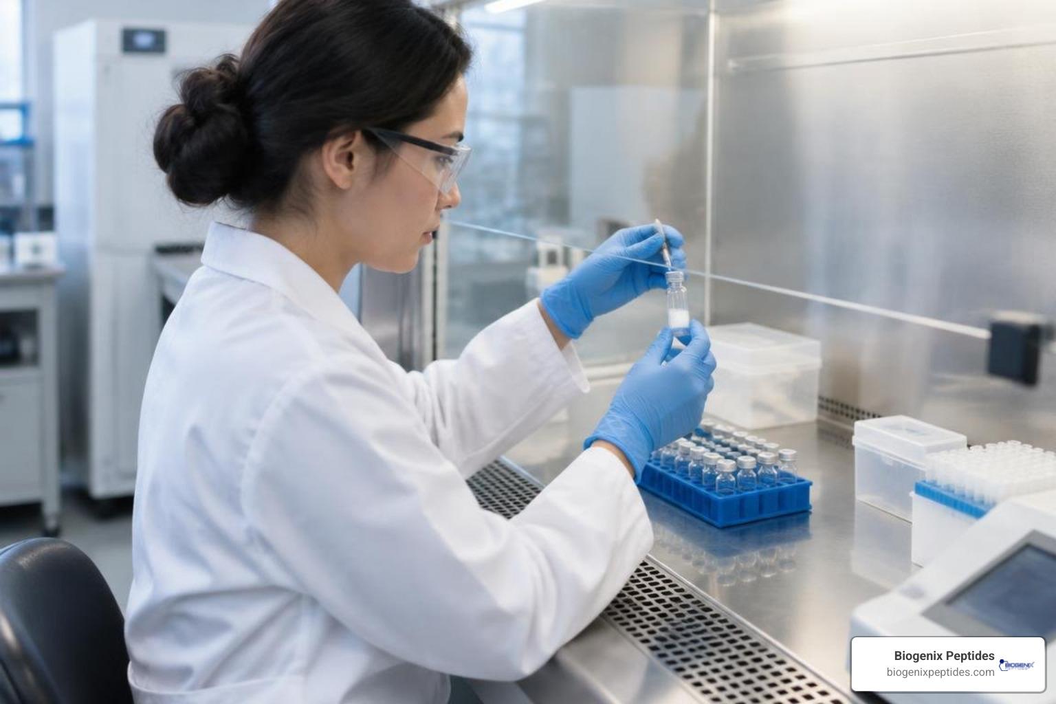
How to Reconstitute Lyophilized Powder in 4 Easy-to-Follow Steps
Disclaimer: The information provided in this article is for educational and informational purposes only. It is not intended as medical advice, diagnosis, or treatment. Products and compounds referenced are for research use only and are not approved for human or veterinary consumption. Always consult a qualified professional regarding health or medical decisions.
How to Use Lyophilized Powder for Research
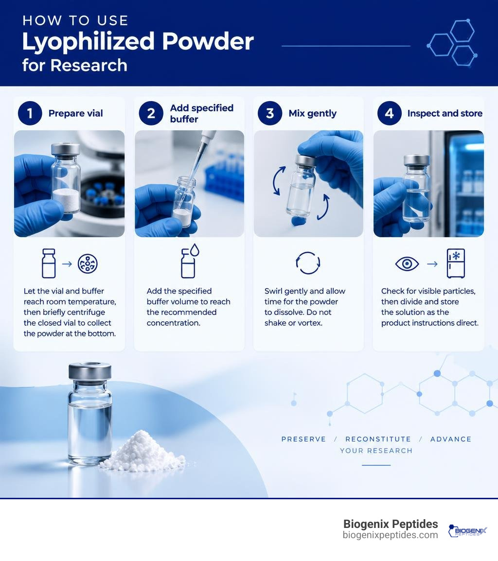
To understand how to use lyophilized powder in a research lab, follow the product instructions and take four steps:
- Let the vial and buffer reach room temperature, then briefly centrifuge the closed vial to collect the powder at the bottom.
- Add the specified buffer volume to reach the recommended concentration.
- Swirl gently and allow time for the powder to dissolve. Do not shake or vortex.
- Check for visible particles, then divide and store the solution as the product instructions direct.
These steps help protect the protein and keep your research samples consistent. I’m Jay Daniel, founder of BioGenix Peptides; my work in peptide sourcing and quality control shapes how I explain how to use lyophilized powder for research. Before the full process, it helps to know why these materials are freeze-dried.
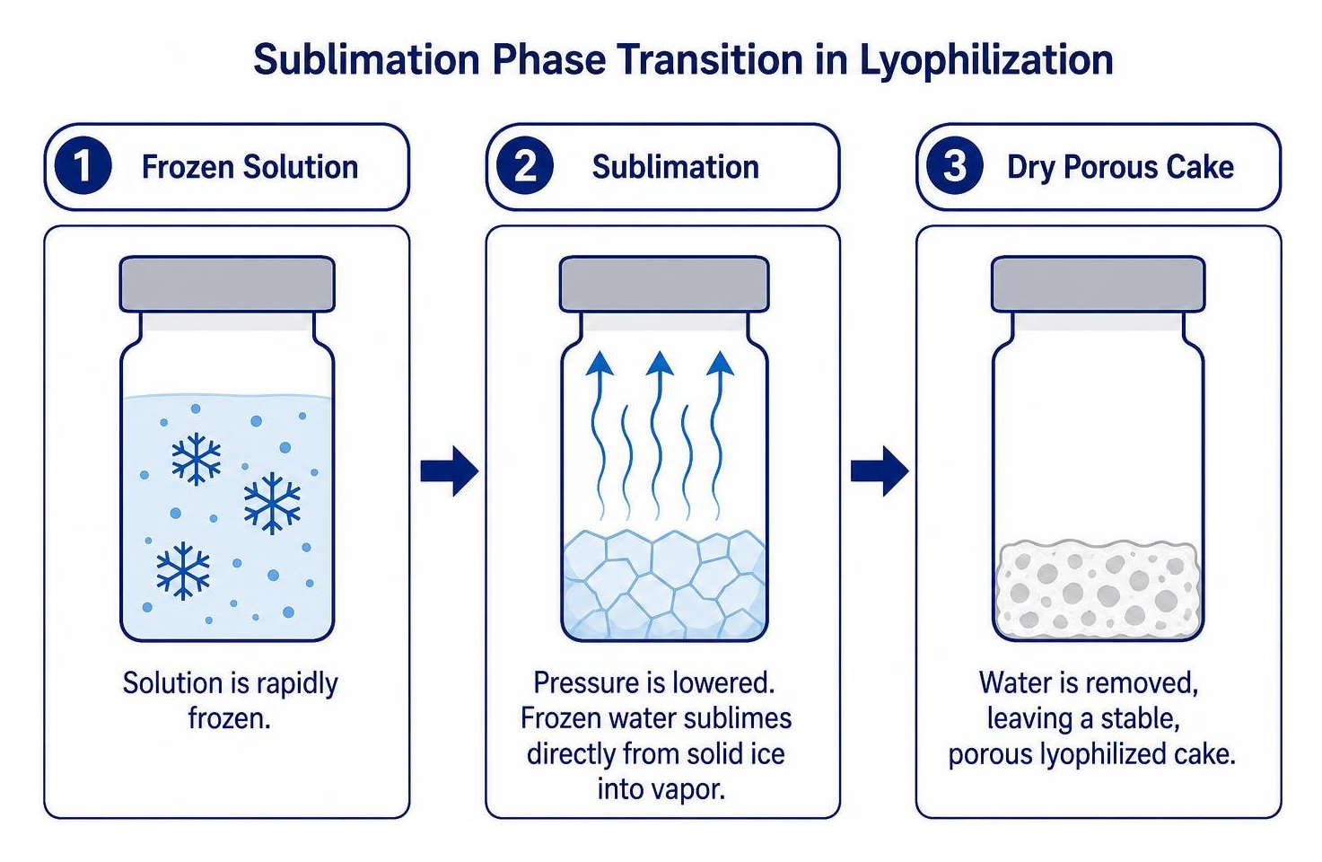
Understanding Lyophilization: Why Proteins and Peptides Are Freeze-Dried
In laboratory research, handling sensitive biological compounds requires methods that preserve molecular integrity over long periods. Lyophilization, commonly known as freeze-drying, is the gold standard for preserving delicate macromolecules, recombinant proteins, and synthetic research chains. Liquid solutions are inherently vulnerable to hydrolytic degradation, enzymatic breakdown (proteolysis), and temperature-induced denaturation.
During the freeze-drying process, a formulated solution is rapidly frozen, and the surrounding pressure is lowered to allow frozen water within the material to sublime directly from solid ice into vapor. This leaves behind a stable, porous structure often referred to as a lyophilized cake.
Removing water effectively pauses molecular movement and chemical degradation pathways. Understanding why research peptides are typically lyophilized and how this benefits research highlights several critical advantages for laboratory teams:
- Extended Shelf Life: Lyophilized cakes can remain stable at room temperature or standard refrigeration for prolonged transit times without rapid degradation.
- Protection Against Proteolysis: Without free water molecules, bacterial enzymatic pathways and cleavage reactions cannot proceed.
- Reduced Cold-Chain Dependency: Solid powders tolerate minor shipping temperature fluctuations far better than hydrated solutions.
- Preserved Bioactivity: When properly freeze-dried with appropriate excipients, secondary and tertiary molecular structures remain preserved upon reconstitution.
How to Use Lyophilized Powder: 4-Step Reconstitution Protocol
Working with freeze-dried materials requires attention to sterile technique and gentle physical handling. Before starting your bench work, ensure your workspace is clean by following standard procedures for creating a sterile research environment.
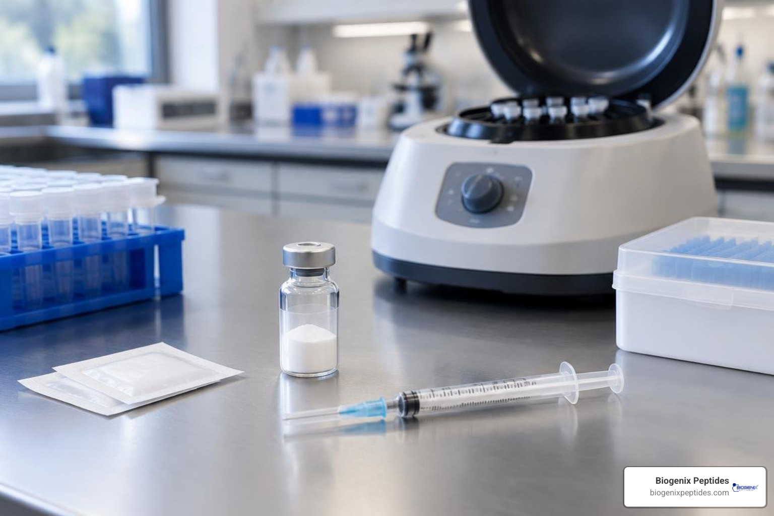
Gather the necessary laboratory supplies:
- Unopened vial of lyophilized powder
- Specified sterile diluent or reconstitution buffer
- Sterile laboratory pipettes or transfer syringes
- 70% isopropyl alcohol prep pads
- Microcentrifuge (or benchtop mini-centrifuge)
- Sterile polypropylene microcentrifuge tubes for aliquoting
Mastering how to mix lyophilized powder reconstitute in simple steps ensures you avoid denaturing delicate molecules during hydration. Most standard laboratory protocols for research proteins recommend a target working concentration between 0.1 mg/mL and 1.0 mg/mL to maintain optimal solubility and molecular stability.
Step 1: Centrifuge and Equilibrate the Lyophilized Vial
Lyophilized material is light, electrostatic, and can shift during shipping. Fine particles often cling to the rubber stopper or upper walls of the glass vial. Opening or piercing the vial immediately can lead to sample loss.
- Thermal Equilibration: Allow both the lyophilized vial and the diluent to sit at controlled laboratory room temperature (20°C to 25°C) for 15 to 20 minutes. Introducing cold diluent to cold powder can slow dissolution and create thermal shock.
- Centrifugation: Place the sealed vial into a centrifuge and spin at 3,000 to 3,500 rpm for 5 minutes. If a specialized vial centrifuge is unavailable, a brief spin in a tabletop unit or firm tapping on a padded benchtop will help consolidate the dry pellet at the base of the glass.
- Sanitization: Wipe the top of the rubber stopper thoroughly with a fresh 70% isopropyl alcohol wipe and allow it to air-dry completely before proceeding.
Step 2: Selecting Diluents: How to Use Lyophilized Powder with Proper Buffer Solutions
Choosing the correct diluent is critical for successful reconstitution. The chemical composition, pH, and ionic strength of your liquid phase dictate whether the freeze-dried material dissolves completely or precipitates into insoluble aggregates.
To calculate the exact volume of buffer needed for your desired stock concentration:
Diluent Volume (mL) = Total Mass of Powder (mg) ÷ Desired Target Concentration (mg/mL)
For instance, to reconstitute a 2 mg vial of research material to a target stock concentration of 0.5 mg/mL, calculate 2 divided by 0.5, yielding 4.0 mL of sterile buffer. Using our practical guide on the science of the bacteriostatic water peptide ratio helps streamline these laboratory calculations.
| Diluent Type | pH / Composition | Primary Laboratory Applications | Key Handling Notes |
|---|---|---|---|
| Bacteriostatic Water | Sterile water with 0.9% benzyl alcohol | Multi-access laboratory research vials requiring antimicrobial protection | Preserves sterility across multiple entries; store at 2°C–8°C after opening. |
| Sterile Water (SWFI) | Pure, non-pyrogenic sterile water | Single-use experimental assays; highly sensitive in-vitro cell cultures | No antimicrobial agent; must be used immediately or frozen into aliquots. |
| Phosphate-Buffered Saline (PBS) | Isotonic phosphate buffer, pH 7.2–7.4 | Cell culture assays, binding kinetics, physiological pH assays | Verify that the specific powder formulation does not precipitate in high phosphate. |
| Dilute Acid/Base (e.g., 10–50 mM Acetic Acid) | Low or high pH aqueous solutions | Hydrophobic or basic sequences that aggregate in neutral pH | Use only when specified on the manufacturer’s technical data sheet. |
Step 3: Gentle Buffer Addition and Dissolution
Adding liquid to a freeze-dried cake requires care. Many research vials contain an internal vacuum, which can draw in liquid rapidly if a needle or pipette tip punctures the septum directly over the pellet.
- Aspirate Buffer: Draw the precisely calculated volume of diluent into a sterile syringe or precision pipette.
- Angle the Stream: Insert the needle through the sanitized rubber stopper and direct the stream against the inside glass wall of the vial rather than directly onto the cake. Let the liquid slide down the glass.
- Release Vacuum Gently: If drawing liquid with a syringe, allow the internal negative pressure to pull the plunger slowly, keeping your thumb on the plunger to prevent sudden splashing.
- Gentle Dissolution: Never shake or vortex the vial. Vigorous mechanical agitation creates hydrodynamic shear stress, which can unravel tertiary molecular folds and form irreversible fibrils. Instead, gently roll the vial between your palms or swirl it slowly in horizontal circles for 30 to 60 seconds.
Step 4: Resolving Particulates: How to Use Lyophilized Powder When Flakes Remain
After gentle swirling, inspect the vial under good laboratory lighting against a dark and light background. The solution should appear uniform and clear.
If visible flakes, floating particulates, or slight turbidity persist, do not discard the sample. Many complex proteins require extra time to fully rehydrate. Reviewing how to reconstitute peptides safely and avoid costly mistakes can help prevent premature sample loss.
- Room-Temperature Incubation: Let the vial rest undisturbed on the laboratory bench for 15 to 30 minutes at room temperature (20°C–25°C).
- Low-Speed Agitation: If flakes remain, place the vial on a slow laboratory rocker or orbital platform shaker set to minimal speed for an additional 15 minutes.
- Refrigerated Dissolution: For stubborn particulate matter, transfer the sealed vial to a refrigerator set at 2°C to 8°C overnight. The extended hydration period at low temperature allows dense molecular clusters to dissolve without thermal degradation.
Storage and Stability Standards for Reconstituted Solutions
Once reconstituted into a liquid state, research compounds become susceptible to degradation pathways. Establishing proper storage protocols is essential to preserve experimental reproducibility. Understanding the definitive guide to how long reconstituted peptides last and referencing the definitive guide to properly storing peptides provide clear frameworks for laboratory storage.
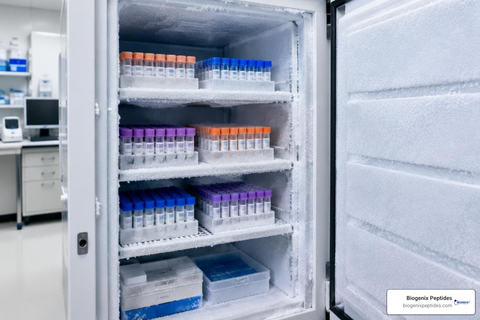
- Short-Term Storage (2°C to 8°C): Reconstituted protein solutions without antimicrobial agents should be kept refrigerated at 2°C to 8°C for no more than 7 days. If reconstituted in bacteriostatic diluents, stability can extend longer depending on the molecule’s chemical nature.
- Long-Term Cryopreservation (-20°C to -80°C): For storage spanning several months, solutions must be divided into working aliquots and frozen at -20°C or -80°C.
Carrier Proteins and Cryoprotection for Long-Term Storage
At low concentrations (e.g., less than 0.1 mg/mL), proteins and peptides often adhere to the hydrophobic surfaces of glass vials or polypropylene microcentrifuge tubes through surface adsorption. This non-specific binding can significantly deplete the active concentration of your working stock.
To prevent adsorption and protect molecular structures during freezing:
- Add Carrier Proteins: Dilute your working stock in a buffer containing a carrier protein, such as 0.1% Bovine Serum Albumin (BSA), 10% Fetal Bovine Serum (FBS), or 5% Human Serum Albumin (HSA). The carrier protein saturates non-specific binding sites on tube walls, keeping your target molecule in solution.
- Cryoprotectants: Adding trehalose or glycerol (5% to 50% concentration) stabilizes protein hydration shells and prevents damaging ice crystal formation during freezing.
- Single-Use Aliquots: Divide your stock solution into single-experiment volumes (greater than 20 µL per vial). Repeated freeze-thaw cycles cause cryo-concentration, pH shifts, and mechanical shearing from growing ice crystals.
Veterinary and Clinical Considerations: Lessons from Fuzapladib Sodium
Commercial lyophilized formulations in veterinary research offer valuable insights into reconstitution stability and handling. A clear example is the investigational veterinary compound fuzapladib sodium (marketed as PANOQUELL®-CA1 for canine pancreatic research models).
According to veterinary product data from the DailyMed – PANOQUELL CA1- fuzapladib sodium, powder, lyophilized, for solution monograph:
- Each vial contains 14 mg of lyophilized fuzapladib sodium.
- The powder is reconstituted using precisely 3.5 mL of the supplied dedicated sterile diluent, yielding an active concentration of 4 mg/mL.
- Unlike fragile research peptides that degrade rapidly in liquid, this stabilized commercial formulation remains stable under refrigeration at 2°C to 8°C for up to 28 days post-reconstitution.
This demonstrates how tailored buffers, controlled ionic environments, and specialized excipients can substantially extend the functional lifespan of reconstituted powders.
Frequently Asked Questions About Lyophilized Powder Reconstitution
Why should you avoid vortexing or shaking reconstituted solutions?
Vortexing and vigorous shaking introduce violent liquid-air interfaces and high shear forces. These mechanical stresses disrupt delicate non-covalent interactions (hydrogen bonds and hydrophobic packing) that hold protein structures together. Denatured molecules expose their hydrophobic interiors, leading to irreversible aggregation, cloudiness, and complete loss of bioactivity. Always mix through gentle swirling, slow inversion, or low-speed rocking.
How long does a reconstituted protein or peptide solution remain stable?
Unpreserved aqueous solutions stored under standard refrigeration (2°C to 8°C) should be used within 7 days. Commercial formulations with protective excipients (such as fuzapladib sodium) can remain stable for up to 28 days at 2°C to 8°C. For extended storage over several months, store samples at -20°C to -80°C in single-use aliquots containing a carrier protein (such as 0.1% BSA).
What should you do if the powder fails to dissolve completely after adding diluent?
If flakes or cloudy precipitates remain after initial mixing:
- Allow the closed vial to sit undisturbed at room temperature for 15 to 30 minutes to facilitate slow rehydration.
- Gently invert the vial three to four times (do not shake).
- Place the vial in a refrigerator at 2°C to 8°C overnight; low-temperature hydration often resolves difficult aggregates.
- Confirm that the diluent pH matches the product data sheet recommendations, as extreme pH shifts can cause precipitation.
Conclusion
Understanding the proper techniques for handling lyophilized powder ensures your laboratory investigations remain reliable, accurate, and reproducible. By allowing vials to equilibrate to room temperature, consolidating powders through brief centrifugation, introducing buffers gently along the vial wall, and avoiding mechanical shear stress, you preserve the structural integrity of sensitive research compounds.
Whenever working with specialized research proteins, always maintain strict sterile handling standards and store reconstituted solutions in single-use aliquots to avoid freeze-thaw damage. To refine your laboratory techniques and expand your understanding of reconstitution methods, explore our comprehensive guide on reconstituting lyophilized peptides step by step.