
The Complete Guide to Peptide Blends for Muscle Growth
Disclaimer: The information provided in this article is for educational and informational purposes only. It is not intended as medical advice, diagnosis, or treatment. Products and compounds referenced are for research use only and are not approved for human or veterinary consumption. Always consult a qualified professional regarding health or medical decisions.
What Research Says About Peptide Blends for Muscle Growth
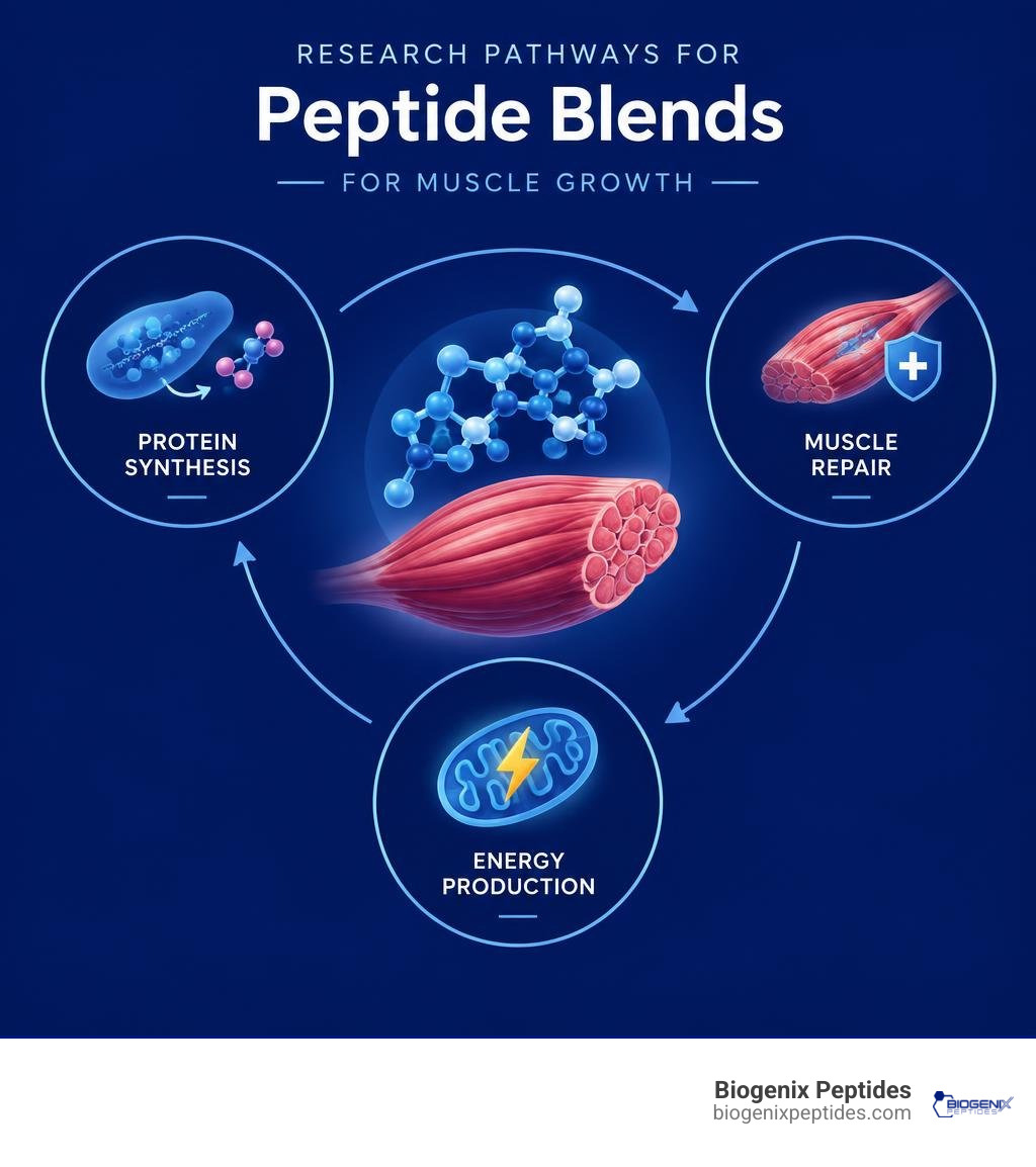
Peptide blends for muscle growth are combinations researchers study for their potential to affect more than one muscle pathway, such as protein synthesis, tissue repair, and recovery. But a plausible mechanism is not proof that a blend builds muscle or improves strength. For now, resistance training and adequate protein have much stronger evidence for supporting muscle health.
That distinction matters because muscle loss is not just a problem of size. With age, muscles can also lose strength as nerve-to-muscle signaling becomes less reliable. Research on long COVID likewise shows that poor exercise capacity can come with changes in muscle fibers and energy production. These findings offer useful questions for peptide research, not proven answers.
I’m Jay Daniel, founder and CEO of BioGenix Peptides, where my work focuses on peptide research, sourcing, and quality standards. In this guide, I’ll examine the evidence behind peptide blends for muscle growth and where the science still falls short.
Cellular Mechanisms of Peptide Blends for Muscle Growth

Skeletal muscle represents approximately 42% of total body mass in an average adult male and 36% in an average adult female. Beyond generating force for locomotion, muscle functions as an active endocrine organ and the body’s primary metabolic reservoir for amino acids.
At the cellular level, muscle hypertrophy relies on the balance between muscle protein synthesis (MPS) and muscle protein breakdown (MPB). Signaling cascades governing this equilibrium depend heavily on the mechanistic target of rapamycin complex 1 (mTORC1) pathway. When stimulated by mechanical tension, amino acids, or anabolic endocrine factors, mTORC1 phosphorylates downstream effectors such as p70S6 kinase and 4E-BP1, accelerating the translation of contractile proteins like actin and myosin.
Simultaneously, myogenic stem cells—known as satellite cells—reside between the basal lamina and sarcolemma of myofibers. In response to cellular stress or structural microtrauma, satellite cells activate, proliferate, and fuse with existing fibers, donating their nuclei to maintain transcriptional capacity across expanding sarcoplasmic volumes.
When evaluating multi-peptide systems, understanding peptide blend synergies helps illustrate how divergent molecular pathways can be stimulated concurrently. While single-target agents may modulate only growth hormone release or local perfusion, combined research models explore how concurrent receptor engagement impacts downstream satellite cell dynamics, cellular transcription, and bioelectrical impedance metrics such as the skeletal muscle mass index (SMI).
Overcoming Sarcopenia and Age-Related Muscle Loss
Sarcopenia is characterized by the progressive, involuntary loss of skeletal muscle mass, structural quality, and contractile force. Epidemiological analyses demonstrate that the prevalence of class I sarcopenia reaches roughly 59% in older women compared to 45% in older men, while severe class II sarcopenia affects approximately 10% of older women and 7% of older men (Janssen et al., 2002). In fact, nearly half of adults older than 80 experience significant sarcopenic decline. The clinical consequences are pronounced: the likelihood of functional impairment and physical disability is roughly two times higher in older men and three times higher in older women presenting with class II sarcopenia relative to age-matched controls with a normal SMI (Janssen et al., 2002).
Muscle tissue exhibits a standard physical density of roughly 1.06 kg/L (compared to 0.92 kg/L for adipose tissue). In sarcopenic states, myosteatosis—the infiltration of inter- and intra-muscular fat—diminishes functional muscle density.
Furthermore, aging triggers selective denervation and preferential atrophy of fast-twitch Type IIx and Type IIa fibers, which produce high anaerobic force output. Contemporary clinical research on muscle optimization investigates how targeted peptide signaling might counteract anabolic resistance and attenuate the degenerative loss of high-threshold motor units.
Neuromuscular Junction Integrity and Electrophysiological Signaling
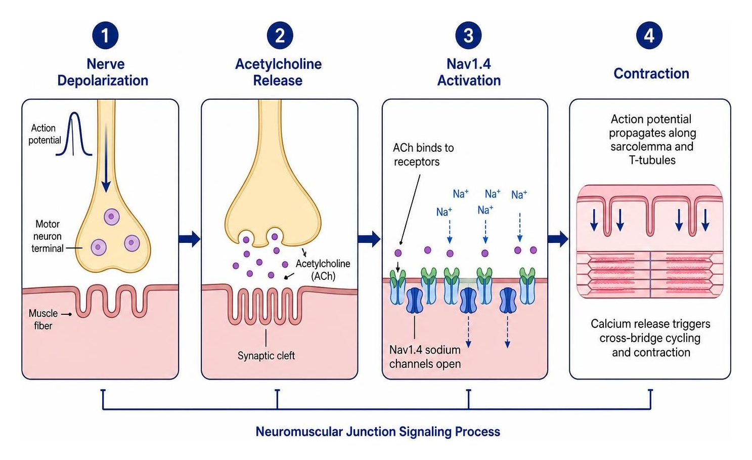
Loss of contractile strength often precedes measurable muscle atrophy, pointing toward a failure in nerve-to-muscle communication. The neuromuscular junction (NMJ) bridges motor neurons and muscle fibers across the motor endplate. Excitation-contraction coupling depends on precise electrophysiological signaling: acetylcholine release triggers endplate potentials, depolarizing the sarcolemma via voltage-gated sodium channels.
Recent investigations highlight the critical role of NaV1.4, a primary voltage-gated sodium channel clustered at the postsynaptic subneural apparatus. With advancing age, localized loss of NaV1.4 decreases endplate potential amplitude and impairs action potential propagation along the T-tubule system.
Concurrently, the skeletal muscle chloride channel ClC-1 stabilizes resting membrane conductance. Emerging preclinical research demonstrates that partial ClC-1 inhibition restores membrane excitability and enhances contractile force generation when NMJ safety margins are compromised. Designing peptide sequences capable of stabilizing NaV1.4 clustering or modulating ion channel kinetics offers an intriguing frontier for supporting neuromuscular transmission and contractile strength.
Metabolic Recovery, Mitochondrial Health, and Tissue Regeneration

Muscle performance and recovery depend on cellular bioenergetics. Recent clinical studies exploring post-exertional malaise (PEM) in complex post-viral syndromes provide critical insights into skeletal muscle vulnerability. In patients experiencing severe exercise intolerance, maximal oxygen uptake (V̇O2max) and peak power output were found to be substantially lower than in healthy cohorts ($p < 0.0001$ and $p = 0.001$, respectively).
Biopsy analyses revealed pronounced metabolic dysfunction, including a significantly lower oxidative phosphorylation (OXPHOS) capacity ($p = 0.003$) and blunted succinate dehydrogenase (SDH) enzymatic activity following exertion. Rather than maintaining an oxidative, fatigue-resistant phenotype, affected tissues exhibited a higher proportion of fast-fatigable glycolytic fibers ($p = 0.036$).
Moreover, post-exertional biopsies demonstrated exercise-induced myopathy characterized by focal myofiber necrosis and extracellular amyloid-containing deposits that increased significantly after physical stress ($p = 0.008$). These findings indicate that compromised mitochondrial respiration and extracellular protein aggregation exacerbate cellular fatigue and structural breakdown, highlighting key pathways for regenerative peptide models to target.
Optimizing Hypertrophy and Recovery: Why Combine Peptide Blends for Muscle Growth?
Relying on a single compound often engages only one pathway, which can lead to receptor saturation or limited adaptive responses across diverse tissue types. In contrast, multi-peptide models investigate how combining distinct mechanisms can simultaneously support microvascular perfusion, mitochondrial recovery, and structural protein synthesis.
| Research Characteristic | Single-Peptide Monotherapy | Multi-Peptide Synergistic Blend |
|---|---|---|
| Primary Pathway Targeted | Isolated receptor cascade (e.g., GH release alone) | Multi-target (secretagogue + cytoprotective + bioenergetic) |
| Receptor Desensitization Risk | Higher if single pathway is chronically stimulated | Lower due to distributed downstream signaling |
| Connective Tissue Support | Minimal (primarily myofibrillar synthesis) | Enhanced (concurrent tenocyte and collagen modulation) |
| Mitochondrial & Metabolic Impact | Indirect | Direct support for cellular respiration and microvascular flow |
| Structural Microtrauma Repair | Standard baseline recovery kinetics | Accelerated extracellular matrix remodeling |
Integrating cytoprotective compounds like BPC-157 for tissue recovery alongside advanced muscle growth and performance peptides allows researchers to study structural microtrauma repair concurrently with anabolic endocrine stimulation. This multi-system approach aims to mitigate exercise-induced myopathy while preserving cellular oxidative capacity.
Growth Hormone Secretagogues and Anabolic Signaling Pathways
The endocrine axis regulating muscle anabolism centers on the pulsatile secretion of growth hormone (GH) from the anterior pituitary, which subsequently stimulates hepatic and peripheral insulin-like growth factor 1 (IGF-1) expression. Rather than introducing exogenous hormones that suppress endogenous endocrine loops, secretagogues stimulate natural release patterns.
A foundational model in peptide research combines growth hormone-releasing hormone (GHRH) analogues with growth hormone secretagogues (GHS / ghrelin receptor agonists). Research on growth hormone secretagogue mechanisms demonstrates how GHRH analogues, such as CJC-1295 (Tetrasubstituted GRF 1-29), bind to pituitary GHRH receptors to increase cyclic adenosine monophosphate (cAMP) levels, recruiting GH-producing somatotrophs.
Concurrently, selective ghrelin mimetics like Ipamorelin stimulate the growth hormone secretagogue receptor (GHSR-1a), activating the phospholipase C (PLC) pathway to trigger intracellular calcium release.
When studied together, this combination produces an amplified, physiological GH pulse without significantly elevating unwanted hormones like prolactin or cortisol. Detailed models, such as clinically guided peptide protocols, examine how sustained IGF-1 expression drives satellite cell activation and systemic nitrogen retention.
Synergistic Protocols and Clinical Assessment Strategies
Peptide signaling pathways operate within a broader physiological network. In research settings, biochemical signaling requires external stimuli—such as progressive overload and essential amino acid availability—to translate into measurable myofibrillar protein synthesis.
Physical loading creates mechanical tension that opens mechanosensitive ion channels and recruits focal adhesion kinase (FAK), directly priming the mTOR pathway. Without adequate protein (typically 0.8 to 1.0 grams per pound of body weight in active models), peptide-mediated signaling lacks the necessary substrate for building contractile tissue.
This consideration is especially relevant in metabolic models involving modern glucagon-like peptide-1 (GLP-1) receptor agonists, where rapid body weight reduction can accelerate the loss of lean tissue alongside adipose mass, making muscle-preserving interventions a key focus of ongoing study.
Clinical Evaluation: Measuring Efficacy of Peptide Blends for Muscle Growth
Objective evaluation of peptide protocols requires tracking functional and systemic biological markers over time:
- Functional Power and Cardiorespiratory Metrics: Measuring peak power output and maximal oxygen uptake (V̇O2max) via incremental cardiopulmonary exercise testing to assess mitochondrial and cardiovascular adaptations.
- Body Composition Analysis: Utilizing dual-energy X-ray absorptiometry (DEXA) or multi-frequency bioelectrical impedance analysis to track changes in skeletal muscle mass index (SMI) and lean tissue mass.
- Serum Endocrine Markers: Monitoring total and free IGF-1, IGFBP-3, and growth hormone pulsatility profiles to verify target receptor engagement.
- Circulating Muscle Damage Indicators: Assessing serum creatine kinase (CK) and myoglobin levels at 24-, 48-, and 72-hour post-exertion intervals to evaluate structural membrane integrity and microtrauma recovery.
- Inflammatory Cytokine Profiling: Measuring high-sensitivity C-reactive protein (hs-CRP), interleukin-6 (IL-6), and tumor necrosis factor-alpha (TNF-$alpha$) to quantify systemic inflammatory responses and recovery kinetics.
Designing Multi-Modal Stacks for Functional Longevity
Comprehensive research protocols explore multi-target systems that combine secretagogues with mitochondrial and cytoprotective peptides. When integrating therapeutic peptide blends, investigators assess how distinct compounds support deep-wave sleep architecture, maintain cellular redox balance, and preserve neuromuscular junction transmission. Aligning mechanical loading, amino acid availability, and targeted peptide signaling provides a structured framework for exploring long-term muscle preservation and physical resilience.
Frequently Asked Questions About Peptide Blends
How do peptide blends differ from single-peptide protocols for muscle development?
Single-peptide protocols stimulate an isolated biochemical receptor, which can result in ceiling effects or receptor desensitization over time. Peptide blends combine compounds that act on complementary biological pathways—such as pairing an extracellular matrix repair peptide with a pituitary secretagogue. This multi-target approach engages both structural healing and systemic protein synthesis pathways simultaneously.
Can peptide blends counteract muscle wasting associated with aging or chronic illness?
In preclinical and clinical research models, specific peptide combinations have been shown to stimulate local protein synthesis, stabilize neuromuscular junction signaling, and support mitochondrial respiration. While these findings highlight promising therapeutic mechanisms for counteracting sarcopenia and post-viral muscle dysfunction, peptide blends remain experimental research tools and are not approved treatments for medical conditions.
What biomarkers best track the effectiveness of a peptide regimen?
The most reliable markers for evaluating peptide efficacy in research include serum IGF-1 levels, skeletal muscle mass index (SMI) via DEXA or bioelectrical impedance, peak force production (such as dynamometer grip strength), and post-exercise creatine kinase clearance rates.
Conclusion
At BioGenix Peptides, our focus is on advancing scientific understanding through rigorous, high-purity formulations designed for advanced laboratory research. Understanding the complex mechanisms of muscle loss—from sarcopenic denervation to post-viral mitochondrial impairment—is essential for developing targeted research models that explore long-term musculoskeletal health.
To learn more about the latest research on tissue repair, mitochondrial support, and cellular regeneration, explore our scientific library of therapeutic recovery peptide solutions.
References
Janssen I, Heymsfield SB, Ross R. “Low relative skeletal muscle mass (sarcopenia) in older persons is associated with functional impairment and physical disability..” Journal of the American Geriatrics Society, 2002. PMID 12028177.