Stop the Clump with the Best Amyloid Aggregation Study Services
Disclaimer: The information provided in this article is for educational and informational purposes only. It is not intended as medical advice, diagnosis, or treatment. Products and compounds referenced are for research use only and are not approved for human or veterinary consumption. Always consult a qualified professional regarding health or medical decisions.
Why Reliable Amyloid Peptide Aggregation Studies Matter
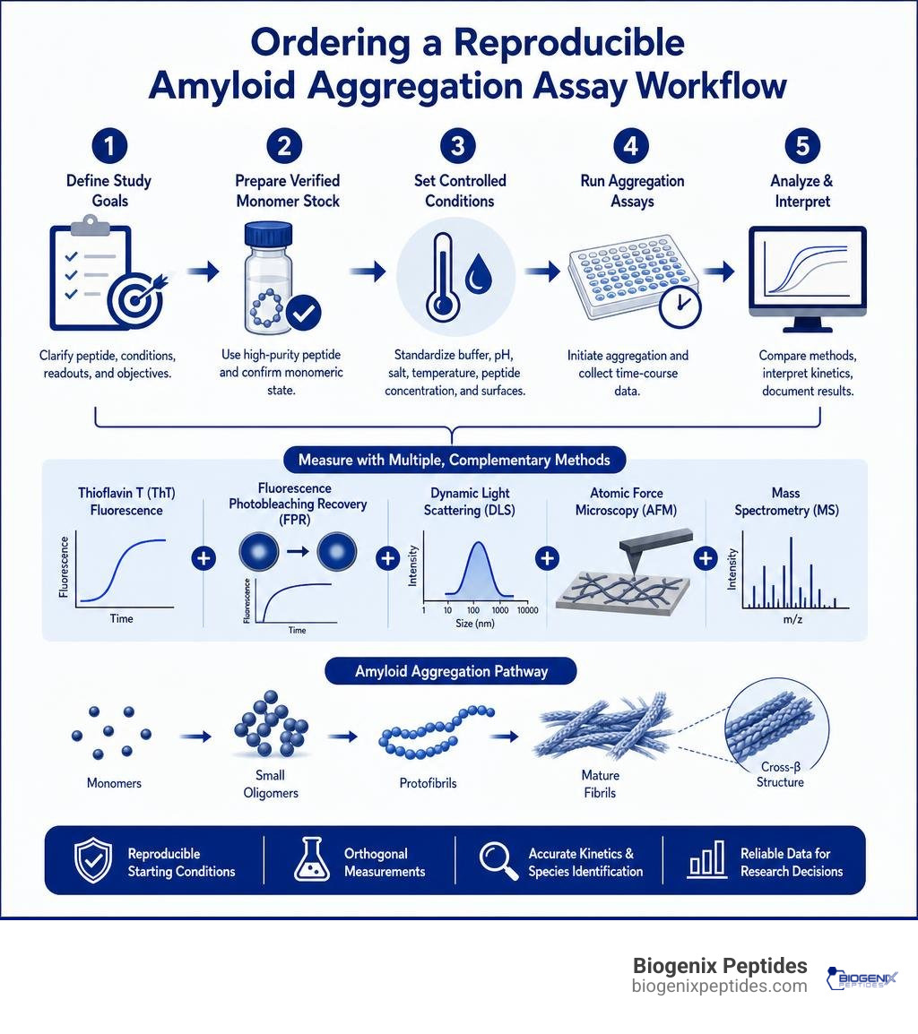
To order useful amyloid peptide aggregation studies, request a standardized, research-only workflow that begins with a verified monomeric peptide stock, tightly controls buffer conditions, and measures aggregation with more than one method. Pair a kinetic assay such as Thioflavin T fluorescence with an orthogonal technique, such as fluorescence photobleaching recovery (FPR), dynamic light scattering (DLS), atomic force microscopy, or mass spectrometry, so early oligomers and later fibrils are not confused.
Aβ peptides, most commonly Aβ1-40 and Aβ1-42, can shift from monomers to small oligomers, protofibrils, and mature cross-beta fibrils. Those transitions are highly sensitive to pH, salt level, temperature, peptide concentration, surfaces, and pre-existing seeds. A clean starting condition is therefore essential for reproducible inhibitor screening and aggregation-kinetics research.
FPR can be especially valuable when small oligomers coexist with large aggregates, because DLS strongly favors the scattering signal from larger particles and dust. Carefully designed studies can also test how peptide mediators, lipid bilayers, or pressure treatment change diffusion, fibril growth, and aggregate shape.
I am Jay Daniel, Founder and CEO of BioGenix Peptides, with experience in peptide quality, sourcing, purity validation, and research-focused laboratory support. My work in amyloid peptide aggregation studies centers on helping researchers build more consistent starting conditions and choose practical analytical workflows.
Biophysical Methodologies in Amyloid Peptide Aggregation Studies

When working with peptides in scientific research, tracking the self-assembly of amyloidogenic sequences requires a multi-faceted biophysical toolkit. The self-association of the amyloid-β (Aβ) peptide—derived via endoproteolytic cleavage of amyloid precursor protein (APP) into fragments typically spanning 39 to 43 amino acids—spans multiple orders of spatial and temporal magnitude. Relying on a single analytical readout frequently leads to erroneous kinetic conclusions.
Standard research workflows incorporate classic tinctorial assays, such as Thioflavin T (ThT) binding kinetics, to monitor beta-sheet enrichment. However, ThT fluorescence predominantly detects cross-beta architecture found in mature fibrils and late protofibrils, remaining virtually blind to low-order, non-fibrillar oligomers. Complementary structural techniques are essential:
- Circular Dichroism (CD) Spectroscopy: Tracks the macroscopic transition from random coil (disordered monomer) to characteristic β-sheet minima (~218 nm).
- Atomic Force Microscopy (AFM): Directly visualizes morphology, differentiating spherical pre-fibrillar assemblies, protofibrillar chains, and mature unbranched fibrils (~10 nm in diameter).
- Native Ion Mobility Mass Spectrometry (IM-MS): Resolves low-abundance, transient oligomeric states by measuring collision cross sections (CCS), distinguishing compact solution-like conformations from extended oligomers without ensemble averaging.
| Biophysical Technique | Primary Readout | Strengths | Limitations in Aggregation Studies |
|---|---|---|---|
| Thioflavin T (ThT) | Fluorescence intensity at ~485 nm | High-throughput, real-time kinetic curves | Blind to early non-fibrillar oligomers; dye interference artifacts |
| Atomic Force Microscopy (AFM) | Nanoscale topography & height profiles | Direct visualization of single particles and fibril morphology | Static imaging; requires surface immobilization |
| Dynamic Light Scattering (DLS) | Autocorrelation of scattered light | Non-destructive, tracks overall size distribution | Heavily biased toward large particles (intensity scales with the sixth power of particle radius) |
| Fluorescence Photobleaching Recovery (FPR) | Translational diffusion coefficient | Resolves coexisting small oligomers and large fibrils | Requires validated fluorophore labeling |
| Native IM-MS | Mass-to-charge ratio and CCS | Resolves stoichiometric oligomer populations (from monomers up to octamers and larger) | Complex instrument optimization; gas-phase compaction limits |
Fluorescence Photobleaching Recovery vs Dynamic Light Scattering
A central analytical challenge in amyloid biophysics is quantifying early oligomeric species in solutions containing polydisperse mixtures of monomers, protofibrils, and mature fibrils. Dynamic Light Scattering (DLS) is widely used to obtain hydrodynamic radius distributions, capturing signal dynamics across ten decades of lag time. However, DLS is governed by Rayleigh scattering, where scattering intensity scales with the sixth power of the particle radius. Consequently, trace quantities of large aggregates or ambient dust overpower the signal, effectively masking the presence of dimers, trimers, and low-order oligomers.
Fluorescence Photobleaching Recovery (FPR) directly circumvents this limitation. In FPR, a brief, intense laser pulse bleaches a defined spatial pattern in a fluorescently labeled sample, and the subsequent recovery of fluorescence is monitored as unbleached molecules diffuse back into the interrogation volume. Because FPR measures mass transport via translational diffusion coefficients, signal amplitude is proportional to the concentration of fluorescently tagged species rather than their volume squared. Monomeric Aβ(1-40) exhibits a well-characterized diffusion coefficient of 1.58 × 10⁻⁶ cm²/s.
When coupled with in situ dialysis cells, FPR allows continuous monitoring of diffusion coefficients over extended incubation periods without mechanical agitation or sample dilution, accurately resolving multi-component diffusion decays where DLS reports only an aggregated average.
High Hydrostatic Pressure Disaggregation for Clean Starting States
The primary impediment to reproducible amyloid kinetics is the presence of pre-existing, kinetically trapped “seeds.” Traditional protocols rely on aggressive fluorinated solvents such as hexafluoroisopropanol (HFIP) or alkaline stock dissolution in 10 mM KOH (pH 11) to promote monomerization. High Hydrostatic Pressure (HHP) provides a powerful physical, non-chemical alternative to dissociate preformed aggregates.
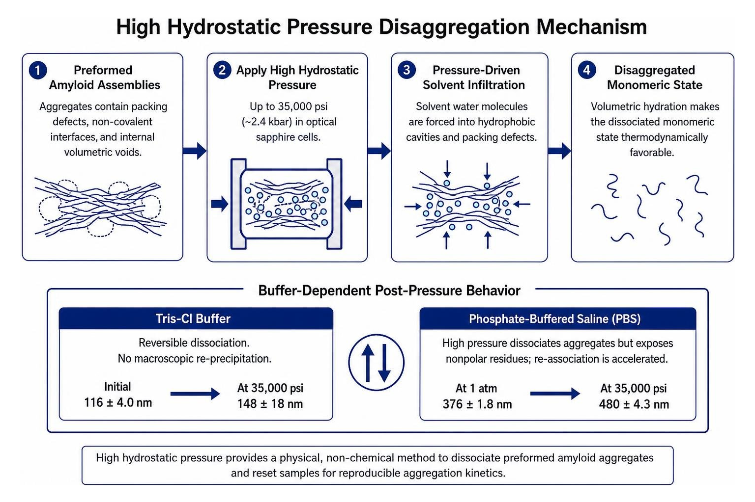
Proteins and amyloid assemblies inherently contain packing defects, non-covalent interfaces, and internal volumetric voids. Application of high hydrostatic pressure (up to 35,000 psi or ~2.4 kbar) in specialized optical sapphire cells forces solvent water molecules into these hydrophobic cavities. This volumetric hydration renders the dissociated monomeric state thermodynamically favorable.
However, buffer composition strongly modulates post-pressure reassociation kinetics:
- In Tris-Cl buffer, preformed Aβ assemblies exhibiting an initial hydrodynamic radius of 116 ± 4.0 nm experience reversible dissociation, stabilizing at 148 ± 18 nm even at 35,000 psi without macroscopic re-precipitation.
- In Phosphate-Buffered Saline (PBS), high pressure dissociates aggregates but simultaneously exposes nonpolar residue clusters; upon release, this hydrophobic exposure can accelerate re-association, driving measured radii from 376 ± 1.8 nm at atmospheric pressure to 480 ± 4.3 nm at elevated pressures.
Kinetic Mechanisms Governing Amyloid Nucleation and Fibril Growth
Amyloid self-assembly is an autocatalytic, non-linear process governed by primary nucleation, fibril elongation, and secondary surface nucleation. Computational and experimental models show that Nucleation and Growth of Amyloid Fibrils follow distinct structural transitions. Primary nucleation initiates in bulk solution via transient oligomeric intermediates where two laminated β-sheets form in a perpendicular orientation before rotating into parallel alignment with the main fibril axis.
Fibril elongation proceeds via a two-stage “dock-and-lock” mechanism. Monomers dock at growing fibril tips either directly from bulk solution or after two-dimensional diffusion along the fibril body, followed by a slower locking phase where side-chain interactions achieve template-directed structural alignment. Furthermore, nonpolar surface patches on existing fibrils serve as catalytic platforms for secondary nucleation, drastically lowering the free-energy barrier for new oligomer generation.
Advancements like a Long-term amyloid aggregation kinetic assay utilizing programmable microdroplet water extraction have demonstrated that compartmentalization alters nucleation stochasticity. By confining aggregating systems to sub-nanoliter droplet volumes, the absolute number of nucleation-capable molecular ensembles is restricted, prolonging lag times and allowing precise deconvolution of stochastic primary nucleation rates from bulk ensemble kinetics.
Structural Role of the Beta-Hairpin in Amyloid Peptide Aggregation Studies
At the monomeric and early oligomeric stage, conformational dynamics dictate the aggregation trajectory. High-resolution native IM-MS and all-atom molecular dynamics indicate that A Hairpin Motif in the Amyloid-β Peptide Is Important for Formation of Disease-Related Oligomers. Intramolecular contact between the central hydrophobic core (CHC, residues 17–21, LVFFA) and the C-terminal hydrophobic region (CTR, residues 30–40) stabilizes a distinct β-hairpin intermediate.
Up to tetrameric states (four peptide units or fewer), oligomers expand isotropically according to a spherical volume scaling law, where the collision cross section of an oligomer containing n units equals the monomer cross section multiplied by n raised to the two-thirds power. As assemblies grow beyond tetramers (more than four units), stabilizing the β-hairpin promotes extended, antiparallel intermolecular β-sheet laminations that drive linear, anisotropic elongation into cytotoxic pre-fibrillar assemblies. Truncating the hydrophobic C-terminus (such as in Aβ1-28) abolishes this second β-strand, arresting self-assembly primarily at dimeric and trimeric stages.
Environmental Factors Modulating In Vitro Peptide Assembly
In vitro amyloid aggregation is acutely sensitive to physical and chemical parameters:
- pH Fluctuations: Near the isoelectric point of Aβ (pH ~5.5), charge neutralization of acidic residues (Glu22, Asp23) minimizes electrostatic repulsion, causing rapid isoelectric precipitation rather than ordered fibrillization.
- Ionic Strength: Elevated salt concentrations shield intermolecular electrostatic repulsion between Lys16 and Glu22, accelerating hydrophobic collapse and primary nucleation rates.
- Temperature & Concentration: Fibrillization displays a critical aggregation concentration (CAC). Elevated temperatures enhance hydrophobic association, shortening nucleation lag times while altering final fibril polymorph distribution.
Advanced Interfacial Dynamics and Inhibitor Screening Protocols
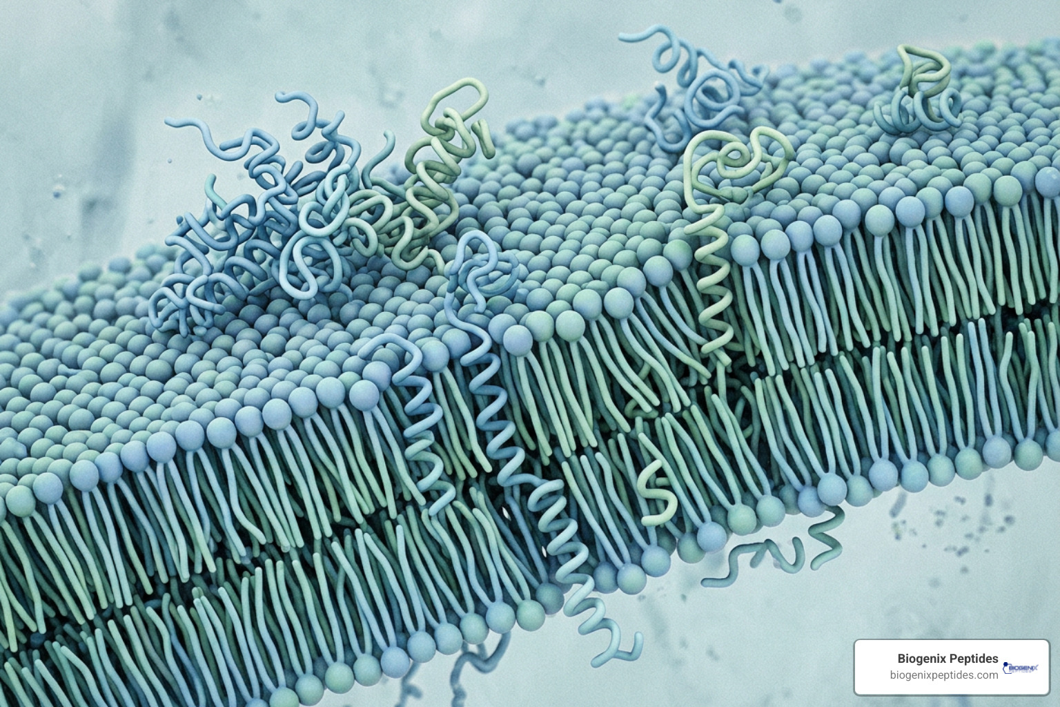
Aggregation kinetics in vivo do not occur in dilute bulk solution; they are catalyzed by macromolecular crowding and cellular interfaces. Amphiphilic Aβ peptides partition strongly at hydrophilic–hydrophobic boundaries, such as outer cell membranes. Confinement to a two-dimensional plane reduces conformational entropy, elevating local peptide density and accelerating β-hairpin nucleation.
When Aβ interacts with supported model phospholipid bilayers (e.g., 1-palmitoyl-2-oleoyl-sn-glycero-3-phosphocholine [POPC] doped with nitrobenzoxadiazole-labeled phosphatidylcholine [NBD-PC]), peptide insertion alters lipid packing. Free NBD-PC/POPC vesicles exhibit a diffusion coefficient of (4.93 ± 0.27) × 10⁻⁸ cm²/s, shifting to (5.99 ± 0.55) × 10⁻⁸ cm²/s upon supported bilayer formation on mica. Membrane insertion of oligomers alters this lateral mobility, reflecting membrane thinning and pore formation linked to cytotoxic mechanisms.
Endogenous chaperones regulate these interfaces. For instance, Apolipoprotein D acts as a chaperone of Aβ peptide delaying its assembly into amyloid aggregates by utilizing its hydrophobic pocket to engage monomeric and oligomeric states at sub-stoichiometric ratios, suppressing structural transitions into β-stranded configurations. Synthetic approaches often mimic this behavior using Short Peptides as Inhibitors of Amyloid Aggregation to cap growing fibril tips.
Screening Peptide-Based Mediators and Modulators
A prominent strategy in anti-amyloid screening involves non-fibrillar peptide-based mediators designed with non-natural α,α-disubstituted amino acids (e.g., Amy1, Amy2). These peptidomimetics disrupt the regular hydrogen bonding network of the cross-beta spine:
- Equimolar Ratios (1:1 Amy2:Aβ): Analysis shows multi-component diffusion profiles, with the largest aggregated species comprising roughly 60% of the total signal amplitude.
- Moderate Excess (2:1 Amy2:Aβ): The system resolves into two diffusing exponentials, with the slower species dominating at 78%.
- High Excess (10:1 Amy2:Aβ): A single diffusing species is observed with a translational diffusion coefficient equivalent to monomeric Aβ (approximately 1.58 × 10⁻⁶ cm²/s), demonstrating complete inhibition of higher-order assembly.
Synthesizing these constrained peptidomimetics requires precision solid-phase protocols, detailed in our peptide synthesis complete guide.
Methodological Quality Control in Amyloid Peptide Aggregation Studies
Assay reproducibility requires rigorous characterization of peptide raw materials and labeled tracers. Chemical impurities, residual trifluoroacetic acid (TFA), or incorrect counter-ions alter aggregation lag times unpredictably.
Researchers must employ rigorous HPLC and mass spectrometry in peptide analysis to confirm sequence fidelity and absolute purity prior to initiating kinetic runs. When fluorophore labeling is necessary (e.g., coupling 5-carboxyfluorescein to the N-terminus), researchers must verify that the modification does not perturb native assembly behavior.
Tracer validation is confirmed by comparing diffusion coefficients of labeled peptide against unlabeled standards via Diffusion-Ordered Spectroscopy (DOSY NMR). Furthermore, reviewing analytical data via guidelines on how to read a peptide COA ensures starting stocks are free of pre-aggregated multimeric fractions.
Frequently Asked Questions About Amyloid Aggregation Assays
Why does dynamic light scattering struggle to quantify small Aβ oligomers?
DLS measures the intensity of scattered light, which is proportional to the particle diameter raised to the sixth power under Rayleigh scattering conditions. In a polydisperse mixture containing both small oligomers (hydrodynamic radius 1–3 nm) and mature fibrils (lengths >200 nm), the light scattered by large aggregates completely overwhelms the signal from smaller oligomers. In contrast, techniques like Fluorescence Photobleaching Recovery (FPR) measure concentration-weighted fluorescence recovery, allowing deconvolution of monomeric and early oligomeric diffusion coefficients without large-particle bias.
How does high hydrostatic pressure prepare monomeric amyloid samples?
High hydrostatic pressure (up to 35,000 psi) exploits the negative volume change associated with the hydration of internal protein cavities. Amyloid fibrils and preformed aggregates contain imperfect internal packing voids and non-covalent interfaces. Elevated pressure forces water molecules into these nonpolar cavities, solvating hydrophobic contact sites and rendering dissociated, monomeric peptide states thermodynamically favorable without the use of harsh chaotropic chemical denaturants.
What steps ensure fluorescent tags do not alter amyloid self-assembly kinetics?
To prevent labeling artifacts, maintain low tracer ratios (typically 1–5% labeled peptide mixed with 95–99% unlabeled native peptide). Conjugate fluorophores strictly to the disordered N-terminus (e.g., Asp1) rather than the central hydrophobic core (residues 17–21) or C-terminal region. Finally, validate that the diffusion coefficient and ThT aggregation kinetics of the tracer mixture match the label-free wild-type peptide using orthogonal DOSY NMR and CD spectroscopy.
Conclusion
Standardizing in vitro amyloid research requires multi-modal biophysical workflows that resolve the full structural spectrum from transient monomers to mature fibrils. By integrating fluorescence photobleaching recovery, dynamic and static light scattering, high hydrostatic pressure disaggregation, and native mass spectrometry, research teams can eliminate seeding artifacts and rigorously quantify aggregation modulators.
BioGenix Peptides delivers premium, analytically validated peptides synthesized under stringent quality control standards for research applications. To ensure optimal experimental starting baselines, review our comprehensive protocol on reconstituting lyophilized peptides step-by-step.
Disclaimer: All products and peptide compounds discussed are intended strictly for in vitro laboratory research and educational use only. They are not intended for human or veterinary use, clinical diagnosis, or medical application.
References
Itoh SG, Okumura H. “Promotion and Inhibition of Amyloid-β Peptide Aggregation: Molecular Dynamics Studies..” International journal of molecular sciences, 2021. PMID 33668406. Finder VH, Glockshuber R. “Amyloid-beta aggregation..” Neuro-degenerative diseases, 2007. PMID 17429215. Khaled M, Rönnbäck I, Ilag LL, Gräslund A, Strodel B, Österlund N. “A Hairpin Motif in the Amyloid-β Peptide Is Important for Formation of Disease-Related Oligomers..” Journal of the American Chemical Society, 2023. PMCID PMC10450692. Jalali S, Zhang R, Haataja MP, Dias CL. “Nucleation and Growth of Amyloid Fibrils..” The journal of physical chemistry. B, 2023. PMCID PMC11426289.