Advances in Longevity | Exploring the Science of Biological Aging
Educational & Research-Only Disclaimer: This article is provided for educational and informational purposes only and discusses theoretical and published scientific research related to metabolic signaling. It is not medical advice and is not intended to diagnose, treat, cure, or prevent any disease. All compounds referenced are intended strictly for laboratory research use and are not for human or animal consumption. Statements have not been evaluated by the U.S. Food and Drug Administration (FDA).
Introduction
Longevity science has moved beyond simplistic “anti-aging” claims and toward a clearer question: why do biological systems lose resilience over time—and which signaling networks coordinate that decline? This is a research-first map of the major aging frameworks and the most commonly studied longevity peptides used as tools to explore mitochondrial function, immune balance, epigenetic timing, tissue repair, and redox homeostasis.
Longevity is a systems problem
Aging is best understood as a gradual loss of coordination across interdependent biological systems. At the cellular level, organisms must constantly balance growth versus maintenance, inflammation versus repair, and energy use versus energy conservation. With time, stress signals accumulate—oxidative stress, DNA damage, inflammatory cytokines, and metabolic overload— and the body shifts from flexible adaptation toward a more rigid, pro-inflammatory, repair-limited state. The practical takeaway from modern geroscience is simple: meaningful longevity signals are rarely “single-target.” Instead, they emerge when multiple systems become more resilient at once.
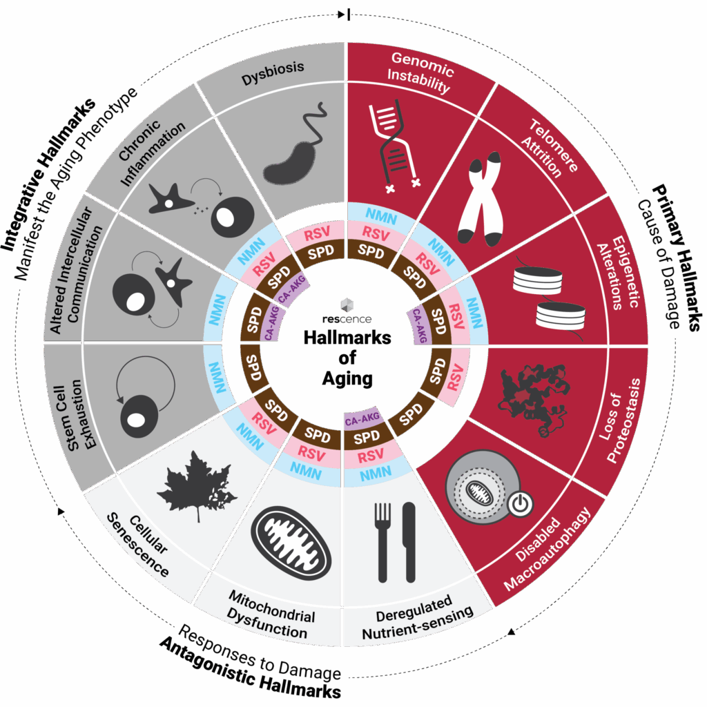
The Hallmarks framework: a map, not a checklist
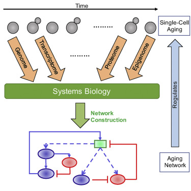
One of the most influential organizing models in longevity research is the “Hallmarks of Aging” framework. Its value is not simply in naming categories (like mitochondrial dysfunction or epigenetic alterations), but in showing how they reinforce one another. Mitochondrial stress can amplify inflammatory signaling; chronic inflammation can accelerate cellular senescence; and senescent cells can further disrupt tissue communication. In other words, aging is a network phenomenon: a change in one node often propagates across the entire system. The most responsible longevity research asks which upstream signals are driving a downstream hallmark, and whether an intervention improves resilience without creating tradeoffs elsewhere.
This page follows that same systems logic. We begin with core biological drivers—mitochondria, redox, nutrient sensing, epigenetic timing, immune tone, and repair capacity—then anchor each domain with the most commonly studied longevity peptides used to explore those pathways.
A simple longevity pathway map (systems view)
The diagram below is a proprietary, simplified “systems map” showing how major longevity domains connect. It is intentionally high-level (education-first) and designed for landing-page clarity rather than biochemical completeness. Mitochondria Energy • redox • stress signaling NAD+ & Redox Repair • sirtuins • oxidative balance Nutrient Sensing AMPK • mTOR • growth/maintenance Epigenetic Timing Gene expression • telomeres • clocks Immune Tone Surveillance • inflammaging • barriers Repair Capacity Angiogenesis • ECM • regeneration Outcome: Higher resilience → slower functional decline Less metabolic overload • better repair • lower chronic inflammation
Mitochondria & cellular resilience
Mitochondria are central to longevity biology because they do far more than produce ATP. They regulate redox signaling, apoptosis, innate immune activation, and metabolic flexibility. With age, mitochondrial populations often show reduced efficiency, impaired turnover (mitophagy), and altered membrane dynamics. Many modern interventions aim to study whether improving mitochondrial signaling and membrane stability can preserve function in high-demand tissues like muscle, heart, and brain.
SS-31 (Elamipretide)
SS-31 (Elamipretide) is a tiny lab-made peptide designed to go straight to your cells’ mitochondria—the parts that make energy. Once there, it attaches to cardiolipin (a key fat in the mitochondrial membrane) and helps keep the mitochondria working more smoothly, which may support steadier energy output and less “wear-and-tear” stress inside the cell—especially in tissues that age hard or work hard (like muscle, heart, and brain).
What makes SS-31 different is that it works inside the mitochondria, not by turning on receptors on the cell surface like many peptides do. Because of that, researchers study it as a tool for understanding mitochondrial health in areas like energy metabolism, heart function, brain resilience, and muscle performance, where mitochondrial stability matters a lot.
MOTS-c
MOTS-c is a tiny peptide that your mitochondria can make. Think of it like a “message molecule” that helps mitochondria communicate with the rest of the cell—especially the nucleus, which controls a lot of gene activity.
When the cell is under metabolic stress (like low fuel or higher oxidative stress), MOTS-c has been observed to move toward the nucleus and help adjust gene expression in ways that support metabolic adaptation and stress response. In simple terms: it’s studied as part of the system that helps cells shift gears and stay resilient when energy conditions get tougher.
Humanin
Humanin is a small, naturally occurring peptide made by the mitochondria, the parts of cells that produce energy. Researchers are interested in Humanin because it appears to help protect cells when they’re under stress and support normal energy balance and cell survival.
Early research suggests Humanin may help cells cope with oxidative stress, support healthier mitochondrial function, and influence pathways tied to aging, metabolism, and resilience. It has also been explored in studies related to brain health, insulin sensitivity, and overall cellular protection.
Humanin belongs to a group called mitochondrial-derived peptides, which act like internal signaling messengers. In simple terms, these peptides help cells sense stress and adjust their behavior to maintain normal function when conditions become challenging.
NAD+ & redox homeostasis
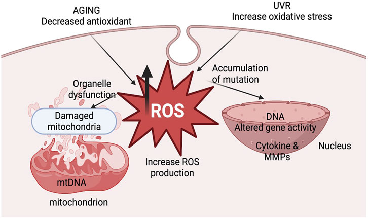
NAD+ sits at the intersection of energy metabolism, DNA repair, and epigenetic regulation. A consistent theme in geroscience is that NAD+ dynamics change with age, influenced by shifts in synthesis, consumption (e.g., PARPs, CD38), inflammation, and tissue composition. Redox imbalance also becomes more likely over time, contributing to oxidative stress and impaired signaling.
Longevity molecule included in this domain: Glutathione (research tool for redox homeostasis). While glutathione is not an “anti-aging treatment,” it is a core molecule in aging biology because shifts in the GSH/GSSG redox state are a common feature of aging tissues.
Nutrient sensing: AMPK & mTOR
Cells continuously decide whether to allocate resources to growth or maintenance. AMPK and mTOR are two central regulators of this balance. AMPK generally supports survival under low-energy conditions by promoting catabolic programs and limiting growth, while mTOR integrates nutrient and hormonal signals to coordinate biosynthesis and anabolic commitment. Longevity research often focuses on flexibility—the ability to shift appropriately between growth and repair—rather than permanent up- or down-regulation of any single pathway.
Epigenetics, telomeres & biological timing
Epigenetic drift—age-related changes in gene expression regulation—tracks strongly with biological aging. In parallel, telomere biology intersects with replicative capacity and cellular stress history. Some ultrashort peptides are studied as “bioregulators,” with hypotheses centered on gene-expression modulation and cellular timing signals (including circadian and pineal-associated coordination). This domain is where longevity research often becomes misunderstood: serious work focuses on mechanisms and model systems, not sweeping “reversal” claims.
Epitalon (Epithalon)
Epithalon (also called Epitalon or Epithalone) is a very small, lab-made peptide made of four amino acids. It’s modeled after substances originally studied in the pineal gland, the part of the brain that helps regulate sleep–wake cycles and biological timing.
In simple terms, researchers study Epithalon because it appears to be involved in cellular “timekeeping”—including circadian rhythm regulation, antioxidant signaling, and how cells age over time. It has been explored in research for its potential links to melatonin regulation, protection against oxidative stress, and pathways related to telomeres and long-term cellular stability.
Because of this connection to biological timing and aging signals, Epithalon is commonly discussed in longevity research focused on sleep regulation, neuroprotection, immune resilience, and age-related cellular decline, rather than short-term stimulation or performance effects.
Pinealon (EDR)
Pinealon is a very small bioregulatory peptide that researchers study for its role in brain health and stress resilience. It’s explored for how it may help brain cells function more smoothly and cope with different kinds of strain, such as physical, emotional, or metabolic stress.
In simple terms, Pinealon is studied for its potential to support clear thinking, memory, and mental adaptability, especially as the brain ages or is under high demand. Because it appears to work by influencing how cells regulate themselves—rather than strongly activating surface receptors—it’s commonly discussed in longevity and neuro-aging research focused on long-term neural stability and resilience.
Immune longevity & “inflammaging”
Aging is accompanied by immune remodeling and a chronic, low-grade inflammatory background often called inflammaging. This inflammatory tone can disrupt metabolic signaling, impair tissue repair, and accelerate functional decline. Longevity research in this domain focuses on maintaining effective immune surveillance while avoiding chronic activation and barrier breakdown.
Thymosin Alpha-1
Thymosin Alpha-1 (Tα1) is a small, naturally occurring peptide linked to the immune system, especially the development and coordination of immune cells. It comes from the thymus gland, an organ that plays a key role in training T-cells, which are central to immune defense.
Researchers study Thymosin Alpha-1 because it appears to help support immune balance and responsiveness, particularly during infection, inflammation, or immune stress. A lab-made version called Thymalfasin is structurally identical to the natural peptide and is produced from a larger precursor protein. In simple terms, Thymosin Alpha-1 is investigated as an immune “signal booster” that may help the body organize and regulate immune responses more effectively under challenging conditions.
LL-37
LL-37 is a naturally occurring peptide that plays an important role in the body’s first line of defense. It is part of the innate immune system and is studied for its ability to help protect the body from bacteria, support healthy inflammation balance, and assist in tissue repair.
In research settings, LL-37 has been shown to interact with harmful microbes, help maintain the strength of the skin and gut barriers, and support the body’s natural healing processes. Studies also explore its potential role in calming irritation, promoting wound recovery, and helping the immune system respond more effectively during stress or injury.
Because LL-37 can influence both immune activity and tissue repair, it is often used as a research tool to better understand how the body protects itself and restores damaged or irritated tissues.
Repair biology & structural integrity
One of the most practical determinants of healthspan is whether repair keeps pace with accumulated micro-damage. Over time, declines in angiogenesis, extracellular matrix (ECM) remodeling, and tissue regeneration can reduce mobility, resilience, and recovery capacity. This domain is where “longevity” becomes measurable in the real world: function, movement, and tissue integrity.
BPC-157
BPC-157 is a small, lab-made peptide originally based on a protective protein found in stomach (gastric) juice. Researchers study it because it appears to play a role in tissue repair and cellular protection, especially in preclinical models.
In simple terms, BPC-157 is explored for how it may help the body repair damaged tissue, support the formation of new blood vessels (angiogenesis), and protect cells under stress. Much of the research focuses on how it interacts with growth-factor signaling pathways involved in healing, making it a common tool for studying wound repair and recovery at the molecular level.
TB-500 (Thymosin β4 fragment)
TB-500 is a lab-made version of Thymosin Beta-4, a protein naturally found throughout the body that’s involved in repair and regeneration. Researchers study TB-500 because it appears to help coordinate tissue healing, especially after physical stress or injury.
In simple terms, TB-500 is explored for how it may support cell movement, blood flow, and the body’s natural repair processes in muscles, tendons, and other soft tissues. Because it’s stable and easy to work with in lab settings, it’s commonly used as a research tool for understanding tissue regeneration and recovery signaling.
Cartalax (AED)
Cartalax is a very small, lab-made peptide made of just four amino acids. Researchers study it because it appears to act as a regulatory signal rather than a typical “on/off” drug that binds to cell receptors.
In simple terms, Cartalax is explored for how it may help cells adjust gene activity related to repair, inflammation, and tissue maintenance—especially in cartilage and joints. Instead of pushing a strong signal from outside the cell, it’s studied for how it may influence the cell’s internal “instructions,” helping tissues respond more normally to stress, wear, and aging.
Because of this, Cartalax is often described as a bioregulatory peptide, and most research interest focuses on connective tissue health, joint integrity, and age-related tissue decline, rather than short-term stimulation effects.
Skin aging, extracellular matrix & antioxidant defense
Skin aging is often treated as cosmetic, but it’s also a readable window into systemic aging biology: collagen remodeling, ECM turnover, wound repair signals, and oxidative stress balance. Two widely discussed longevity-relevant molecules in this domain are GHK-Cu (a copper-binding peptide involved in repair signaling) and glutathione (a foundational redox molecule tied to oxidative defense and signaling fidelity).
GHK-Cu
GHK-Cu is a naturally occurring copper-binding peptide that the body releases at sites of injury. It’s found in blood plasma and plays a role in repair and regeneration signaling, especially in tissues that rely on collagen and structural support.
In simple terms, researchers study GHK-Cu because it appears to help coordinate tissue repair, support healthy collagen production, and assist antioxidant and anti-inflammatory pathways. Levels of GHK-Cu naturally decline with age, which is one reason it’s frequently discussed in aging and skin-biology research. Overall, GHK-Cu is explored as a signaling molecule that helps guide how cells repair, protect, and remodel tissue over time.
L-Glutathione
Glutathione is a small peptide that acts as one of the cell’s main internal antioxidants. It helps protect cells by neutralizing reactive oxygen species and keeping the cell’s internal environment in balance.
In simple terms, glutathione works like a cleanup and protection system inside cells. Researchers often look at the balance between its active form (GSH) and its used form (GSSG) to understand how much oxidative stress a cell is under. Beyond antioxidant defense, glutathione is also studied for its roles in immune regulation, mitochondrial protection, and normal cell survival processes, which is why it’s considered a foundational molecule in aging and longevity research.
Longevity peptide atlas
- SS-31 (Elamipretide) — mitochondrial membrane / cardiolipin research
- MOTS-c — mitochondrial-derived metabolic signaling research
- Humanin — mitochondrial-derived cytoprotection research
- Epitalon (Epithalon) — epigenetic/telomere-associated signaling research
- Pinealon (EDR) — neuro-aging and gene-expression research
- Thymosin Alpha-1 — immune signaling research
- LL-37 — innate defense and barrier signaling research
- BPC-157 — tissue repair / angiogenesis signaling research
- TB-500 — thymosin β4-associated repair signaling research
- Cartalax (AED) — structural longevity / connective tissue gene-regulation research
- GHK-Cu — ECM/collagen and regeneration signaling research
- Glutathione — redox homeostasis research
Peer-review references
References below are provided as peer-review anchors for the educational sections and the specific longevity peptides discussed.
- López-Otín C, Blasco MA, Partridge L, Serrano M, Kroemer G. The hallmarks of aging. Cell. 2013;153(6):1194-1217. PMC • PubMed
- Chavez JD, et al. Mitochondrial protein interaction landscape of SS-31. Proc Natl Acad Sci U S A. 2020. PMC • Publisher
- Mitchell W, et al. The mitochondria-targeted peptide SS-31 binds lipid bilayers and modulates surface electrostatics as a key component of its mechanism. J Biol Chem. 2020. Full text
- Lee C, et al. The mitochondrial-derived peptide MOTS-c promotes metabolic homeostasis and reduces obesity and insulin resistance. Cell Metab. 2015;21(3):443-454. Full text • PubMed
- Lee C, Yen K, Cohen P. Humanin: a harbinger of mitochondrial-derived peptides? Trends Endocrinol Metab. 2013. PMC • PubMed
- Imai S, Guarente L. NAD+ and sirtuins in aging and disease. Trends Cell Biol. 2014. PMC
- Camacho-Pereira J, et al. CD38 dictates age-related NAD decline and mitochondrial dysfunction. Cell Metab. 2016. PMC
- McReynolds MR, Chellappa K, Baur JA. Age-related NAD+ decline. Exp Gerontol. 2020. PMC
- Trefts E, Shaw RJ. AMPK: restoring metabolic homeostasis over space and time. Mol Cell. 2021. PMC
- Kennedy BK, Lamming DW. The mechanistic target of rapamycin: the grand conducTOR of metabolism and aging. Cell Metab. 2016. PMC
- Franceschi C, Garagnani P, Parini P, Giuliani C, Santoro A. Inflammaging: a new immune-metabolic viewpoint for age-related diseases. Nat Rev Endocrinol. 2018. PubMed • Publisher
- Dominari A, Hathaway D, Pandav K, et al. Thymosin alpha 1: a comprehensive review of the literature. World J Virol. 2020. PMC
- Heilborn JD, Nilsson MF, Kratz G, et al. The cathelicidin anti-microbial peptide LL-37 is involved in re-epithelialization of human skin wounds. J Invest Dermatol. 2003;120(3):379-389. PubMed
- Carretero M, et al. In vitro and in vivo wound healing-promoting activities of human cathelicidin LL-37. J Invest Dermatol. 2008. Full text
- McGuire FP, et al. Regeneration or Risk? A narrative review of BPC-157 for musculoskeletal research. 2025. PMC
- Malinda KM, et al. Thymosin β4 accelerates wound healing. J Invest Dermatol. 1999. PubMed
- Xing Y, et al. Progress on the function and application of thymosin β4. Front Mol Biosci. 2021. PMC
- Khavinson VKh, Bondarev IE, Butyugov AA. Epithalon peptide induces telomerase activity and telomere elongation in human somatic cells. Bull Exp Biol Med. 2003;135(6):590-592. PubMed
- Khavinson V, et al. EDR peptide (Pinealon): possible mechanisms of gene expression regulation and neuroprotection. 2020. PMC
- Khavinson VK, et al. Peptide regulation of gene expression: a systematic review. Molecules. 2021. PMC
- Linkova NS, et al. Peptide regulation of chondrogenic stem cell… (AED peptide and connective tissue signaling contexts). Int J Mol Sci. 2023;24(9):8415. Publisher
- Pickart L, Margolina A. Regenerative and protective actions of the GHK-Cu peptide in human skin. Biomed Res Int. 2018. PMC
- Pickart L. GHK peptide as a natural modulator of multiple cellular pathways in skin regeneration and repair. Int J Mol Sci. 2015. PMC
- Aquilano K, Baldelli S, Ciriolo MR. Glutathione: new roles in redox signaling for an old antioxidant. Front Pharmacol. 2014. PMC
- Sekhar RV, et al. Deficient synthesis of glutathione underlies oxidative stress in aging humans (human data). Am J Clin Nutr. 2011. PMC
- Su Y, et al. Progress in understanding how clock genes regulate aging and age-related disease. 2025. PMC